Neurobiology of Propofol Addiction
February 23, 2022
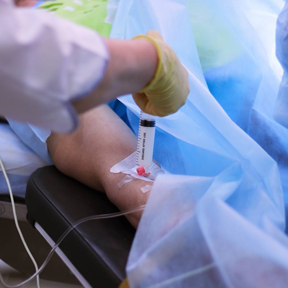
Propofol is a quick-acting, sedating, intravenous anesthetic agent used extensively in surgical and critical care settings. It also has an anti-emetic effect, leading to the design of several postoperative regimens that include propofol to moderate nausea and vomiting. Studies have suggested propofol’s additional anxiolytic and anti-epileptic effects are due to its positive modulation of GABAA receptors [1]. Glycine receptors, which are known to modulate pain perception and motor control, have displayed similar potentiation after propofol administration [2,3]. Patients awaking from propofol anesthesia often report elation or euphoria, and subjects injected with subanesthetic doses of propofol reported feeling high, spaced out, lightheaded, and disoriented, all in a positive sense [4]. Propofol addiction has been reported in numerous case studies worldwide and is thought to be disproportionately present in medical professionals compared to laypeople, due to increased accessibility [5]. A small random-choice experiment in healthy volunteers asked participants to choose between either propofol or placebo after blind sampling of both injections. From a sample size of 12, participants chose propofol at a statistically higher rate than would be expected by chance [4]. These findings highlight an effect of propofol that occurs with other drugs of abuse and can lead to addiction — creating positive experiences that may encourage recreational use. In November 2009, the Drug Enforcement Agency (DEA) classified fospropofol, a prodrug of propofol which has oral bioavailability in the sense that it has an active effect when taken orally, as a controlled substance, a measure supported by the American Society of Anesthesiologists (ASA) and the American Association of Nurse Anesthetists (AANA) [4,6,7,8].
Extracellular signal-regulated kinase (ERK) is central to signal transduction in behavioral output, neuroplasticity, and mood modulation [9]. It is especially abundant in the mesolimbic dopamine system, the neural region of reward circuitry and goal-directed behavior [10]. Recent evidence indicates acute administration of cocaine, nicotine, and morphine increase ERK phosphorylation in the nucleus accumbens (NAcc), a critical terminal area of mesolimbic projections [9]. Wang et al. conducted a study showing rats maintained on propofol had significantly more phosphorylated ERK (p-ERK) in the NAcc and pretreatment with SCH23390, a D1 receptor antagonist, attenuated this p-ERK increase and inhibited propofol self-administration. The study concluded that ERK pathways and D1 dopamine receptors in the NAcc may mediate the addictive component of propofol [9].
The Finkel-Bishin-Jinkins murine osteosarcoma viral oncogene homolog B (FosB) is a transcription factor rapidly induced by addictive drugs [11]. ∆FosB, a truncated form of FosB, features unusual stability in the body, making it likely to mediate long-lasting changes in gene expression long after drug cessation [12]. In preclinical studies, mice expressing greater quantities of ∆FosB work harder to self-administer cocaine, suggesting both increased sensitization to the drug as well as a predisposition towards relapse after withdrawal [12]. Xiong et al. showed both ethanol and nicotine to increase ∆FosB expression; intriguingly, propofol administration yielded similar ∆FosB increases [13]. D1 dopamine receptors were also upregulated, supporting Wang et al.’s suggestion that D1 receptors may play a mediating role in propofol addiction [9,13].
Following previous experiments linking addiction to glucocorticoid functioning, Wang et al. found dexamethasone, a glucocorticoid receptor (GR) agonist, diminished addictive behavior in rats maintained on propofol as well as the expression of D1 receptors in the NAcc [14]. Inhibition of RU486, a GR antagonist, had the same mitigating effect [14].
Propofol shows similarities with different addictive drugs and may lead to similar deleterious consequences. Further exploring the mechanisms of propofol is of vital importance to determining future regulations or caveats on its clinical use.
References
- Trapani, G., Altomare, C., Liso, G., Sanna, E., & Biggio, G. (2000). Propofol in anesthesia. Mechanism of action, structure-activity relationships, and drug delivery. Current Medicinal Chemistry, 7(2), 249–271. https://doi.org/10.2174/0929867003375335
- Lynch, J. W. (2004). Molecular structure and function of the glycine receptor chloride channel. Physiological Reviews, 84(4), 1051–1095. https://doi.org/10.1152/physrev.00042.2003
- Hales, T. G., & Lambert, J. J. (1991). The actions of propofol on inhibitory amino acid receptors of bovine adrenomedullary chromaffin cells and rodent central neurones. British Journal of Pharmacology, 104(3), 619–628. https://doi.org/10.1111/j.1476-5381.1991.tb12479.x
- Zacny, J. P., Lichtor, J. L., Thompson, W., & Apfelbaum, J. L. (1993). Propofol at a subanesthetic dose may have abuse potential in healthy volunteers. Anesthesia and Analgesia, 77(3), 544–552. https://doi.org/10.1213/00000539-199309000-00020
- Welliver, M. D., Bertrand, A., Garza, J., & Baker, K. (2012). Two new case reports of propofol abuse and a pattern analysis of the literature. International Journal of Advanced Nursing Studies, 1(1), 22–42. https://doi.org/10.14419/ijans.v1i1.27
- 2010—Proposed Rule: Placement of Propofol into Schedule IV. Available online: https://www.deadiversion.usdoj.gov/fed_regs/rules/2010/fr1027.htm
- ASA Supports DEA Proposal to Schedule Propofol—American Society of Anesthesiologists (ASA). Available online: http://www.asahq.org/about-asa/newsroom/news-releases/2010/11/asa-supports-dea-proposal-to-schedule-propofol
- Securing Propofol. Available online: https://www.aana.com/docs/default-source/practice-aana-com-web-documents-(all)/securing-propofol.pdf?sfvrsn=2b0049b1_2
- Wang, B., Yang, X., Sun, A., Xu, L., Wang, S., Lin, W., Lai, M., Zhu, H., Zhou, W., & Lian, Q. (2016). Extracellular signal-regulated kinase in nucleus accumbens mediates propofol self-administration in rats. Neuroscience Bulletin, 32(6), 531–537. https://doi.org/10.1007/s12264-016-0066-1
- Siciliano, C. A., Ferris, M. J., & Jones, S. R. (2015). Cocaine self-administration disrupts mesolimbic dopamine circuit function and attenuates dopaminergic responsiveness to cocaine. European Journal of Neuroscience, 42(4), 2091–2096. https://doi.org/10.1111/ejn.12970
- Ulery, P. G., & Nestler, E. J. (2007). Regulation of ΔFosB transcriptional activity by Ser27 phosphorylation. European Journal of Neuroscience, 25(1), 224–230. https://doi.org/10.1111/j.1460-9568.2006.05262.x
- Nestler, E. J. (2008). Transcriptional mechanisms of addiction: Role of ΔFosB. Philosophical Transactions of the Royal Society B: Biological Sciences, 363(1507), 3245–3255. https://doi.org/10.1098/rstb.2008.0067
- Xiong, M., Li, J., Ye, J. H., & Zhang, C. (2011). Upregulation of deltafosb by propofol in rat nucleus accumbens. Anesthesia and Analgesia, 113(2), 259–264. https://doi.org/10.1213/ANE.0b013e318222af17
- Wu, B., Liang, Y., Dong, Z., Chen, Z., Zhang, G., Lin, W., Wang, S., Wang, B., Ge, R.-S., & Lian, Q. (2016). Glucocorticoid receptor mediated the propofol self-administration by dopamine D1 receptor in nucleus accumbens. Neuroscience, 328, 184–193. https://doi.org/10.1016/j.neuroscience.2016.04.029