Spinal Abnormalities in Neuraxial Anesthesia
July 1, 2024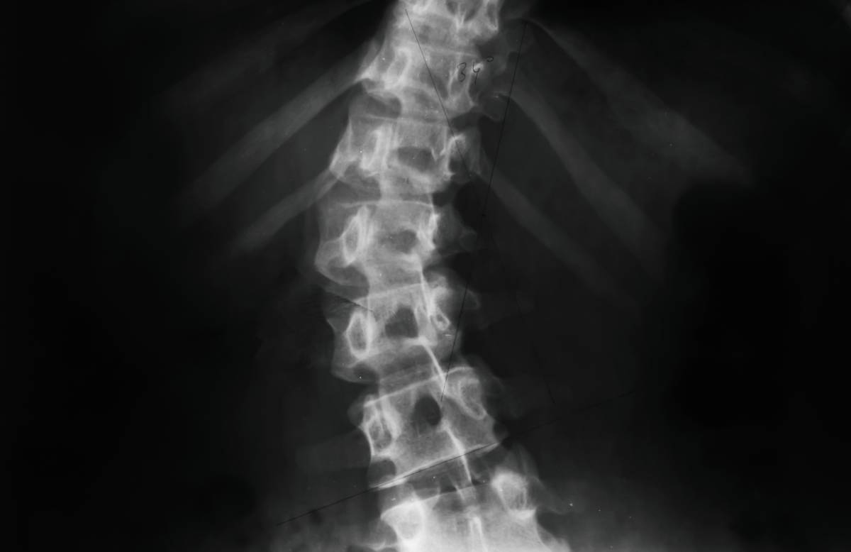
Neuraxial anesthesia refers to a set of techniques which induce analgesia in the abdomen, pelvis, or lower extremities by injecting anesthetic around the spinal cord. Types of neuraxial blockade include spinal anesthesia (injection into the subarachnoid space), epidural anesthesia (injection into the epidural space), and caudal anesthesia (injection near the cauda equina)1. Neuraxial anesthesia has applications in a wide array of clinical scenarios, including patients who are undergoing labor, have underlying respiratory compromise, or are obese2. Although neuraxial anesthesia is an essential tool for the modern anesthesiologist, its use can be challenging when spinal abnormalities are present3. Such abnormalities include scoliosis, lordosis, spina bifida, spinal stenosis, and previous spinal surgery.
Individuals with scoliosis demonstrate abnormal lateral curvature of the spinal column, leading to distortion of the spinous processes and asymmetry of the intervertebral spaces. Abnormalities in spinal curvature and anatomy can increase the difficulty of identifying landmarks and complicate attempts at needle placement for neuraxial anesthesia. Indeed, a 2009 meta-analysis demonstrated that rates of inadequate or failed neuraxial analgesia in obstetric patients with scoliosis were an order of magnitude higher than in patients with normal anatomy4. Exaggerated lordosis, too, can complicate neuraxial anesthesia5 by shifting the spine and epidural space anteriorly.
Spinal canal pathology – such as spinal stenosis, lumbar radiculopathy, lumbar disc disease, and other degenerative changes – can also complicate neuraxial anesthesia. These patients may have decreased space available for needle or epidural catheter insertion and are at an increased risk of neurologic injury following neuraxial blockade compared to the general population6. Patients with a history of spinal surgery may also be relatively difficult candidates for neuraxial techniques due to indwelling hardware, scar tissue, adhesion of the dura to the ligamentum flavum, and/or persistent abnormalities of spinal anatomy8.
Neuraxial anesthesia may also be complicated by congenital vertebral conditions in pediatric and adult populations. Spinal dysraphisms include a broad spectrum of disorders, ranging from spina bifida occulta, in which vertebral fusion is incomplete with few if any clinical findings, to open spinal dysraphisms in which nervous tissue is exposed. Whereas severe spinal dysraphisms are likely to undergo surgical correction, spina bifida occulta frequently goes unnoticed7. Despite surgical correction, severe spinal dysraphisms are often accompanied by persistent neurologic deficits as well as secondary anatomical changes which can confound attempts at needle or catheter placement. For example, patients may demonstrate tethering of the filum terminale, also known as tethered cord syndrome. There is a paucity of data regarding outcomes of neuraxial anesthesia in patients with spinal dysraphisms, particularly for unrepaired, severe defects. The decision to pursue neuraxial blockade must be made on a case-by-case basis depending on the severity of the defect, accompanying neurologic/structural changes, and the ability to pursue needle or catheter insertion without violating the underlying defect7.
When attempting neuraxial anesthesia in patients with spinal abnormalities, it is essential for clinicians to obtain a thorough history and physical exam along with careful review of prior radiologic studies. If needed, further pre-procedural imaging can be helpful in characterizing spinal anatomy. Clinicians may also facilitate needle or catheter insertion by employing periprocedural imaging techniques such as ultrasound or fluoroscopy1,2, or by modifying patient positioning. Even in the presence of spinal abnormalities, selected patients can successfully receive neuraxial anesthesia from a skilled clinician.
References
1. Li, J., Krishna, R., Zhang, Y., Lam, D. & Vadivelu, N. Ultrasound-Guided Neuraxial Anesthesia. Curr. Pain Headache Rep. 24, 59 (2020).
2. Sivakumar, R. K. & Karmakar, M. K. Spinal sonography and central neuraxial blocks. Best Pract. Res. Clin. Anaesthesiol. 37, 209–242 (2023).
3. Walsh, E., Zhang, Y., Madden, H., Lehrich, J. & Leffert, L. Pragmatic approach to neuraxial anesthesia in obstetric patients with disorders of the vertebral column, spinal cord and neuromuscular system. Reg. Anesth. Pain Med. 46, 258–267 (2021).
4. Ko, J. Y. & Leffert, L. R. Clinical implications of neuraxial anesthesia in the parturient with scoliosis. Anesth. Analg. 109, 1930–1934 (2009).
5. Korkmaz Toker, M., Altiparmak, B., Uysal, A. I., Turan, M. & Gumus Demirbilek, S. Rider sitting position widens lumbar intervertebral distance: a prospective observational study. Braz. J. Anesthesiol. Elsevier 73, 758–763 (2023).
6. Hebl, J. R., Horlocker, T. T., Kopp, S. L. & Schroeder, D. R. Neuraxial blockade in patients with preexisting spinal stenosis, lumbar disk disease, or prior spine surgery: efficacy and neurologic complications. Anesth. Analg. 111, 1511–1519 (2010).
7. Chang, L. Y., Carabuena, J. M. & Camann, W. Neurologic issues and obstetric anesthesia. Semin. Neurol. 31, 374–384 (2011).
8. Murphy, C. J., Stanley, E., Kavanagh, E., Lenane, P. E. & McCaul, C. L. Spinal dysraphisms in the parturient: implications for perioperative anaesthetic care and labour analgesia. Int. J. Obstet. Anesth. 24, 252–263 (2015).